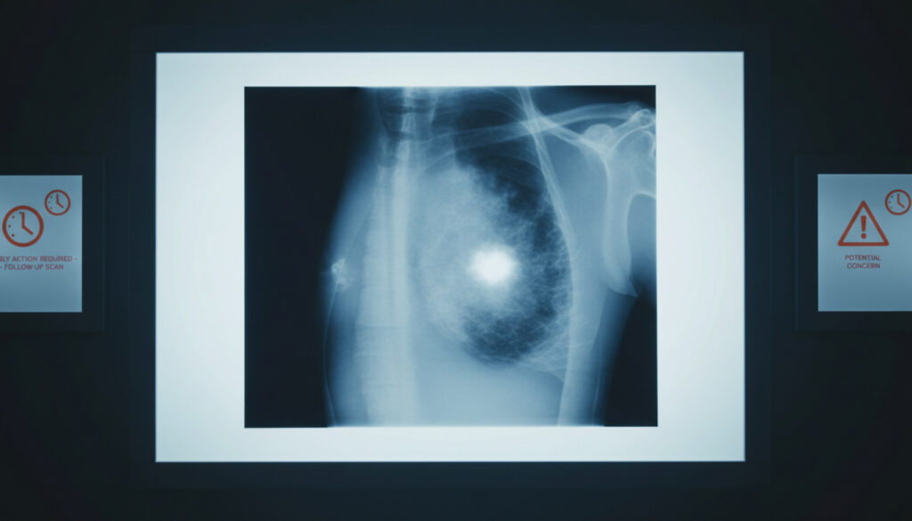
Breast Lump When to Worry – Signs, Causes & When to Act
Discovering a lump in the breast triggers immediate anxiety for most women, yet medical data consistently shows that the majority of these findings are not cancerous. Approximately four out of five breast lumps submitted for biopsy prove benign, arising from conditions such as cysts, fibroadenomas, or fibrocystic changes. Nevertheless, the physical and emotional urgency of the discovery demands prompt attention, because distinguishing between harmless tissue variations and potential malignancy requires professional evaluation rather than self-assessment.
The challenge lies in the variability of symptoms. Benign lumps often present as soft, movable, and sometimes painful masses that fluctuate with hormonal cycles. Cancerous growths, by contrast, typically manifest as hard, irregular, and painless masses that persist or grow over time. Yet exceptions exist in both categories, making physical characteristics alone an unreliable diagnostic tool.
Understanding the specific features that warrant immediate medical consultation helps navigate the gap between normal vigilance and unnecessary panic. This guide examines the tactile qualities of concerning lumps, explores the spectrum of benign causes, and outlines the critical steps for proper evaluation.
What Do Breast Cancer Lumps Feel Like?
Benign Indicators
Soft, movable, often painful masses that may vary with menstrual cycles
Warning Signs
Hard texture, irregular edges, fixed to tissue, typically painless
Immediate Action
Any new, persistent, or changing lump requires prompt GP evaluation
Common Origins
Cysts, fibroadenomas, and hormonal fibrocystic changes
- Most breast lumps—roughly 80%—prove benign upon diagnostic examination
- Cancerous lumps typically feel hard and irregular, though early tumors may remain movable
- Pain does not rule out malignancy, though painful lumps more commonly indicate cysts
- Size alone does not determine risk; even small masses require professional assessment
- Monthly self-exams starting at age 20 establish baseline familiarity with breast tissue
- Upper outer quadrant and armpit locations account for approximately half of cancer presentations
- Changes persisting beyond four to six weeks demand immediate medical imaging
| Feature | Benign Characteristics | Cancer Concern |
|---|---|---|
| Texture | Soft, squishy, or rubbery | Hard, discrete, stone-like |
| Mobility | Movable, slips under skin | Fixed or immobile |
| Pain | Often tender or painful | Usually painless |
| Edges | Smooth, round, defined | Irregular, angular, poorly defined |
| Size/Change | Stable or fluctuates with hormones | Grows or changes over time |
| Location | Anywhere; common in pre-menopause | Often upper outer quadrant or armpit |
Mayo Clinic specialists emphasize that hard texture and irregular edges distinguish potentially malignant masses from benign tissue. Similarly, Mass General Brigham oncologists note that cancerous lumps often feel distinctly different from surrounding breast tissue, presenting as discrete, immovable nodules.
Breast Lump Causes
Breast tissue responds dynamically to hormonal fluctuations, injury, and age-related changes, producing various lump types that differ significantly in origin and risk profile. Understanding these distinctions helps contextualize why most findings do not indicate cancer while highlighting which conditions require closer monitoring.
Fibrocystic Changes and Cysts
Fibrocystic breast changes create a lumpy or rope-like texture throughout the tissue, particularly common among women before menopause. These changes stem from hormonal stimulation and often cause bilateral tenderness that intensifies before menstruation. Cysts—fluid-filled sacs that develop from blocked breast ducts—frequently present as distinct, smooth, mobile lumps that may feel tender or painful. Cleveland Clinic data indicates that cysts represent one of the most frequently encountered benign breast masses in clinical practice.
Fibroadenomas and Other Benign Growths
Fibroadenomas appear as solid, smooth, rubbery tumors most commonly diagnosed in women in their twenties and thirties. Unlike cysts, these fibrous growths typically persist without fluctuating with menstrual cycles. Other non-cancerous causes include fat necrosis following breast injury, lipomas composed of fatty tissue, mastitis infections, and milk retention cysts in breastfeeding women. Certain benign conditions such as atypical hyperplasia or lobular carcinoma in situ slightly elevate future cancer risk despite being non-malignant themselves.
Approximately 80% of breast lumps evaluated through biopsy prove benign, according to the Breast Cancer Research Foundation. This high prevalence reflects the breast’s sensitivity to hormonal and structural changes rather than widespread malignancy.
Breast Cancer How to Check
Systematic self-examination serves as a complementary tool to mammographic screening, helping women recognize normal tissue variations and identify new changes requiring professional evaluation. While self-exams cannot replace imaging technology, they foster body awareness that facilitates earlier detection.
Self-Examination Technique
The examination begins visually. Stand before a mirror with arms at the sides, then raised overhead, and finally pressed firmly on the hips to flex chest muscles. Observe for changes in contour, dimpling, or skin texture. For the manual component, lie supine and use the finger pads—not the tips—of the opposite hand to palpate in circular motions from the outer breast toward the nipple. Vary pressure from light to firm and extend the search to include the armpit and collarbone areas where lymph nodes reside.
Many women find that performing this examination while showering, when skin is slippery, allows for easier movement of fingers over breast tissue. Consistency matters more than exact timing, though experts suggest monthly checks following menstruation when hormones stabilize.
Screening Limitations
Not all breast cancers present as palpable lumps. Research from Ohio State University indicates that many malignancies remain undetectable through touch alone, appearing instead as architectural distortions or calcifications visible only on mammograms. Therefore, women over forty or those with elevated risk factors should maintain scheduled mammographic screening regardless of self-exam findings.
No self-examination technique can definitively distinguish between benign and malignant tissue. Houston Methodist Hospital specialists emphasize that professional evaluation remains essential for any new or persisting mass, as early-stage cancers may feel identical to fibroadenomas during palpation.
Breast Lump When to Worry Reddit
Online forums frequently feature personal accounts of women discovering lumps and seeking reassurance about whether their specific characteristics indicate cancer. While these narratives provide emotional support, they cannot substitute for clinical assessment, particularly regarding long-standing masses or atypical presentations.
Long-Standing Lumps and Stability
Women occasionally report lumps persisting for a decade or longer without significant change. Such stability generally suggests a benign process, particularly if the mass remained unchanged through hormonal shifts, pregnancy, or menopause. However, medical recommendations advise re-evaluation if any new characteristics emerge, including increased size, altered texture, or the onset of pain in a previously painless lump.
The Danger of Visual Diagnosis
Requests for photographic comparisons circulate frequently in online spaces, yet such images prove diagnostically worthless. Stony Brook Medicine clinicians note that breast cancer manifests across a spectrum of physical appearances, and benign conditions can mimic concerning features. CDC guidelines stress that accompanying symptoms—such as skin dimpling, nipple retraction, bloody discharge, or orange-peel texture—warrant immediate attention regardless of the underlying lump’s appearance.
Anecdotes shared on social platforms lack the medical verification necessary for diagnostic purposes. Family history of breast cancer, while elevating risk, does not create certainty about any particular lump’s nature. Only clinical examination combined with appropriate imaging can resolve diagnostic uncertainty.
From First Symptom to Diagnosis
The pathway from discovering a lump to obtaining a definitive diagnosis follows a standardized sequence designed to maximize accuracy while minimizing unnecessary intervention.
- Initial Detection: The woman notices a palpable mass during routine self-exam, showering, or accidental contact while dressing.
- Observation Period: If the lump appeared suddenly or coincides with menstruation, some physicians suggest monitoring for one menstrual cycle, though immediate consultation remains safest for any hard, fixed, or irregular mass.
- Clinical Consultation: The general practitioner performs a physical examination and refers for imaging based on the lump’s characteristics and the patient’s age.
- Diagnostic Imaging: Mammography serves as the primary tool for women over forty, while ultrasound often benefits younger women or those with dense breast tissue.
- Biopsy Procedure: If imaging reveals suspicious features, a needle biopsy extracts tissue samples for histopathological analysis, providing definitive diagnosis.
The fear of serious diagnosis often delays medical visits, yet early detection significantly improves treatment outcomes. Unlike conditions such as stomach ulcers where specific protocols exist for What Is the Fastest Way to Cure a Stomach Ulcer – Healing in 4-8 Weeks, breast lumps require individualized diagnostic pathways rather than standardized cure timelines.
What Doctors Know vs Unknowns
Medical science has established clear parameters for evaluating breast masses, though certain aspects remain uncertain or require individualized interpretation.
| Established Facts | Diagnostic Uncertainties |
|---|---|
| 80% of biopsied lumps prove benign | Exact progression timeline from benign to malignant in high-risk conditions |
| Hard, fixed, painless lumps carry higher cancer probability | Why some painful lumps occasionally represent malignancy |
| Monthly self-exams improve early detection rates | Optimal frequency of imaging for women with dense breast tissue |
| Size changes over 4-6 weeks require evaluation | Significance of lumps stable for decades but suddenly changing |
Context and Risk Factors
Age constitutes the primary risk factor for breast cancer, with incidence rising significantly after fifty. Additional factors include early menstruation onset, late menopause, and family history of breast or ovarian cancer. However, risk factors indicate probability rather than destiny; many women with multiple risk factors never develop cancer, while others with no known risks receive unexpected diagnoses.
Claims regarding “natural cures” for breast lumps through diet, supplements, or topical treatments lack scientific validation. Baystate Health specialists confirm that no dietary modification or alternative therapy provenly eliminates established breast lumps, and pursuing such approaches may delay necessary medical treatment. Similarly, while maintaining adequate Folic Acid Side Effects – Risks, Symptoms and Safety Guide levels supports general cellular health, no vitamin regimen dissolves established masses.
Medical Authority and Sources
Most breast lumps are benign, but any new, persistent, or changing lump requires prompt medical evaluation to rule out breast cancer.
— Breast Cancer Research Foundation Clinical Guidelines
Not all breast cancers start as noticeable lumps. Many are detected through screening before they can be felt.
— Ohio State University Comprehensive Cancer Center
Early-stage cancers may be movable, becoming fixed to surrounding tissue as they progress.
— Mayo Clinic Department of Oncology
Next Steps After Finding a Lump
Schedule an appointment with a general practitioner or breast specialist within days of discovering any new, hard, or irregular lump, particularly if it persists beyond four to six weeks or accompanies skin changes. Bring notes about when you first noticed the mass, whether it changes with your menstrual cycle, and any family history of breast disease. While awaiting your appointment, avoid attempting to manipulate or compress the lump repeatedly, as this may cause tissue irritation. Remember that seeking reassurance through professional channels provides the only reliable path to resolving the uncertainty that accompanies any breast discovery.
Is there a natural cure for breast lumps?
No evidence supports natural cures for breast lumps. Claims involving diet, supplements, or topical treatments remain unproven and may delay necessary medical evaluation. Professional diagnosis through imaging and potential biopsy represents the only reliable approach.
Can I identify cancer by comparing real pictures?
Photographic comparisons cannot reliably distinguish benign from malignant lumps. Breast cancer presents across a physical spectrum, and benign conditions may mimic concerning appearances. Professional clinical assessment and imaging provide necessary diagnostic certainty.
Does a movable lump rule out cancer?
Not entirely. While movable lumps more commonly indicate benign conditions like fibroadenomas or cysts, early-stage cancers may remain mobile before becoming fixed to surrounding tissue. Immobility increases suspicion but does not confirm malignancy.
How urgent is a doctor’s visit for a new lump?
Prompt evaluation within days is advisable for any hard, irregular, or fixed lump, or those persisting beyond four to six weeks. While not typically an emergency, delaying assessment may allow progressive conditions to advance undetected.
Does pain indicate the lump is harmless?
Pain generally suggests benign conditions such as cysts or fibrocystic changes, but does not definitively exclude cancer. Some malignant tumors cause discomfort, making pain an unreliable diagnostic indicator without further testing.
Should I worry about a lump unchanged for ten years?
A decade-long stable mass typically indicates a benign condition like a fibroadenoma. However, any new changes in size, texture, or pain level require re-evaluation, as long-term stability does not guarantee future benign behavior.
More related posts
 Shoes Insoles: Which Type Works Best for You
 Xander Schauffele: Ethnicity, Wife, Children, Net Worth & More
 Puerto Rico: Travel Guide, Facts, Tax Rules, and Pros & Cons
 Is Grok AI Free? Pricing, Features, and ChatGPT Comparison (2025)
 James Gunn: Career, DC Studios Role, and Superman Movie
 Miranda Cosgrove: Relationship, Ethnicity, Injury & Net Worth
 15 Dollars in Pounds – Live Rate and Exchange Guide
 Ruth Gemmell: Biography, Age, Nationality, and Key Roles